Case studies
Each exhibit below was built from the patient's or client's own imaging for a plaintiff firm. Details are anonymized, and every image is a rendering of the exhibit we built.
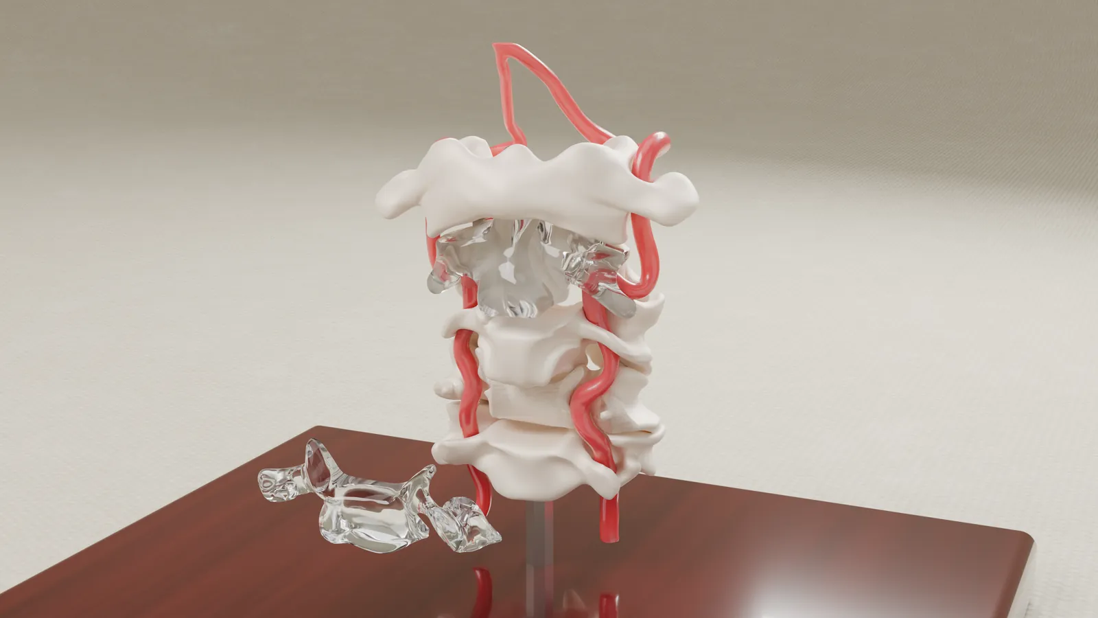
Vertebral artery injury during cervical fusion
A 1:1 cervical spine model and an interactive exhibit, built from the patient's own CT and MRI.
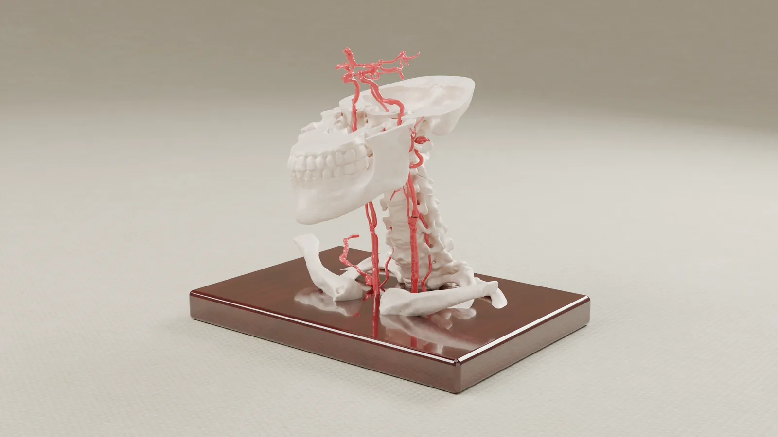
Carotid artery measurement in a wrongful death case
A model of the carotid arteries from the patient's CT angiogram, with the measured section marked in black.
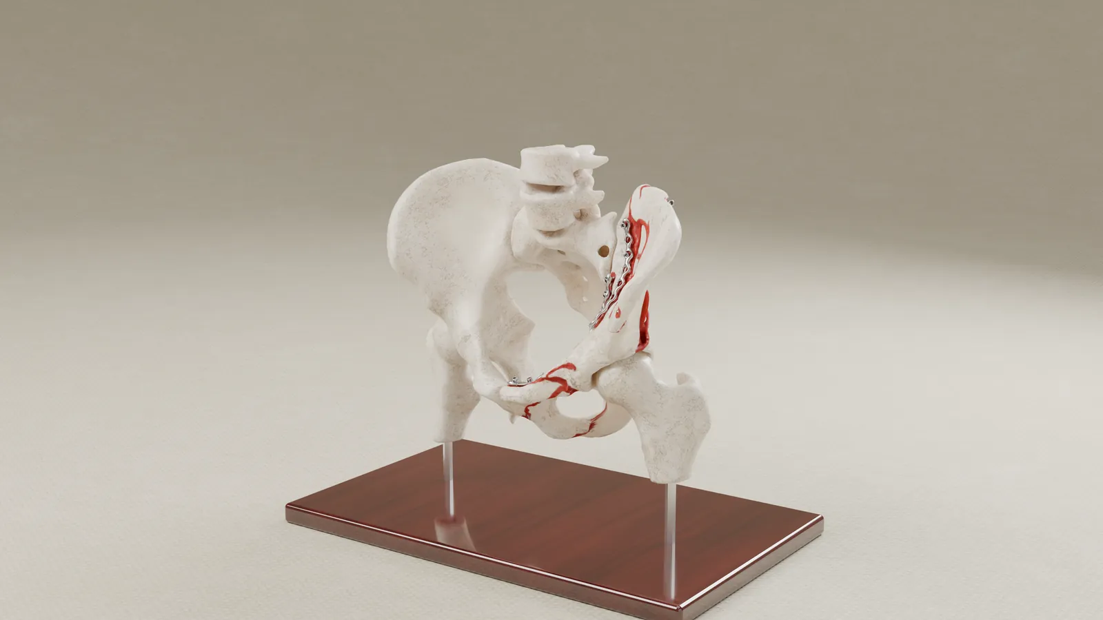
Pelvic fracture and surgical reconstruction
An interactive exhibit that shows the break before surgery and the repair after, from the client's CT scans.
Other exhibits
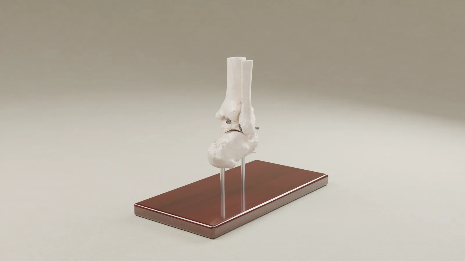
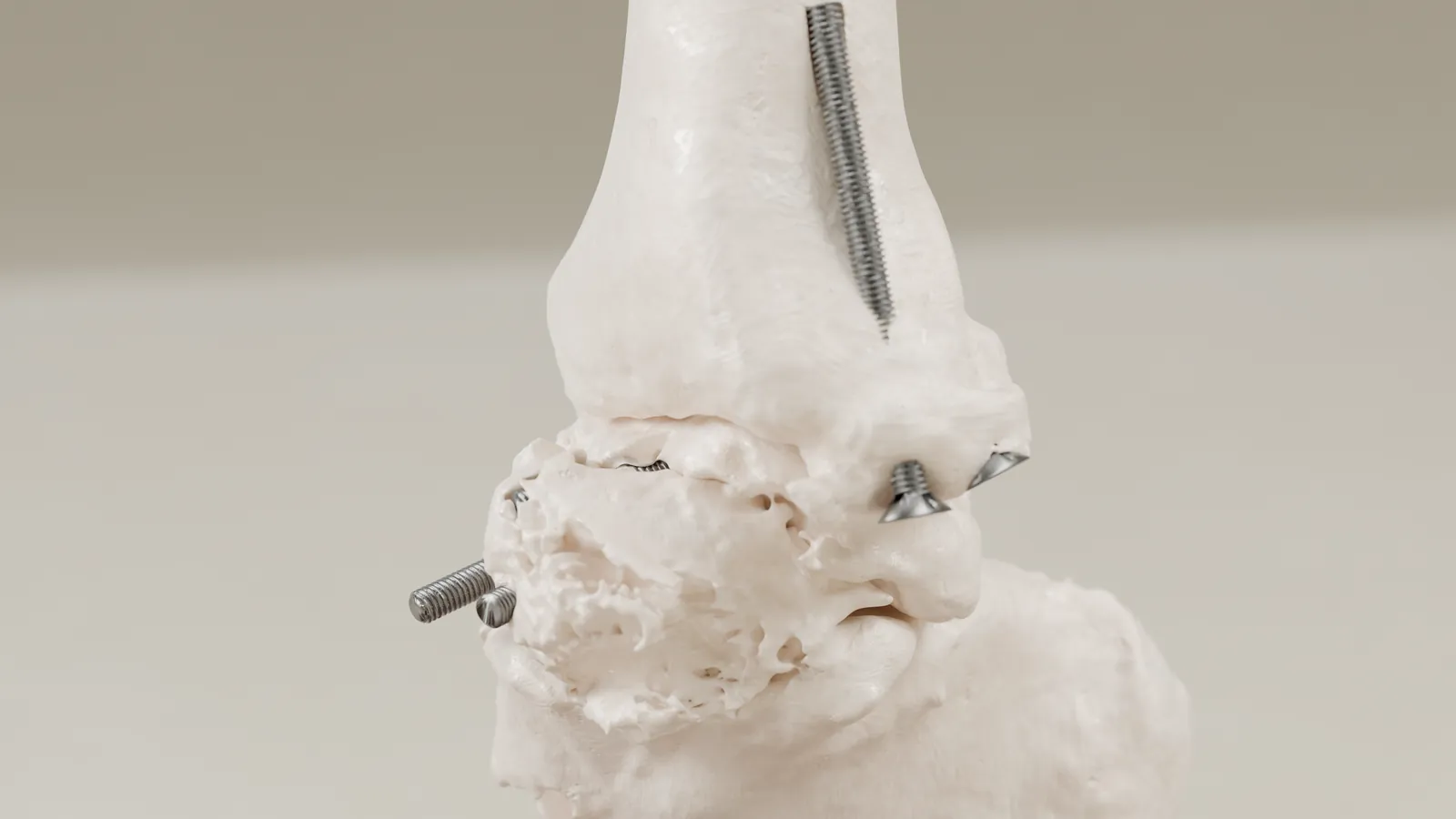
Failed ankle reconstruction
For a failed ankle reconstruction, we built a hand-held model of the bones from the client's own CT, with the screws in their actual positions.
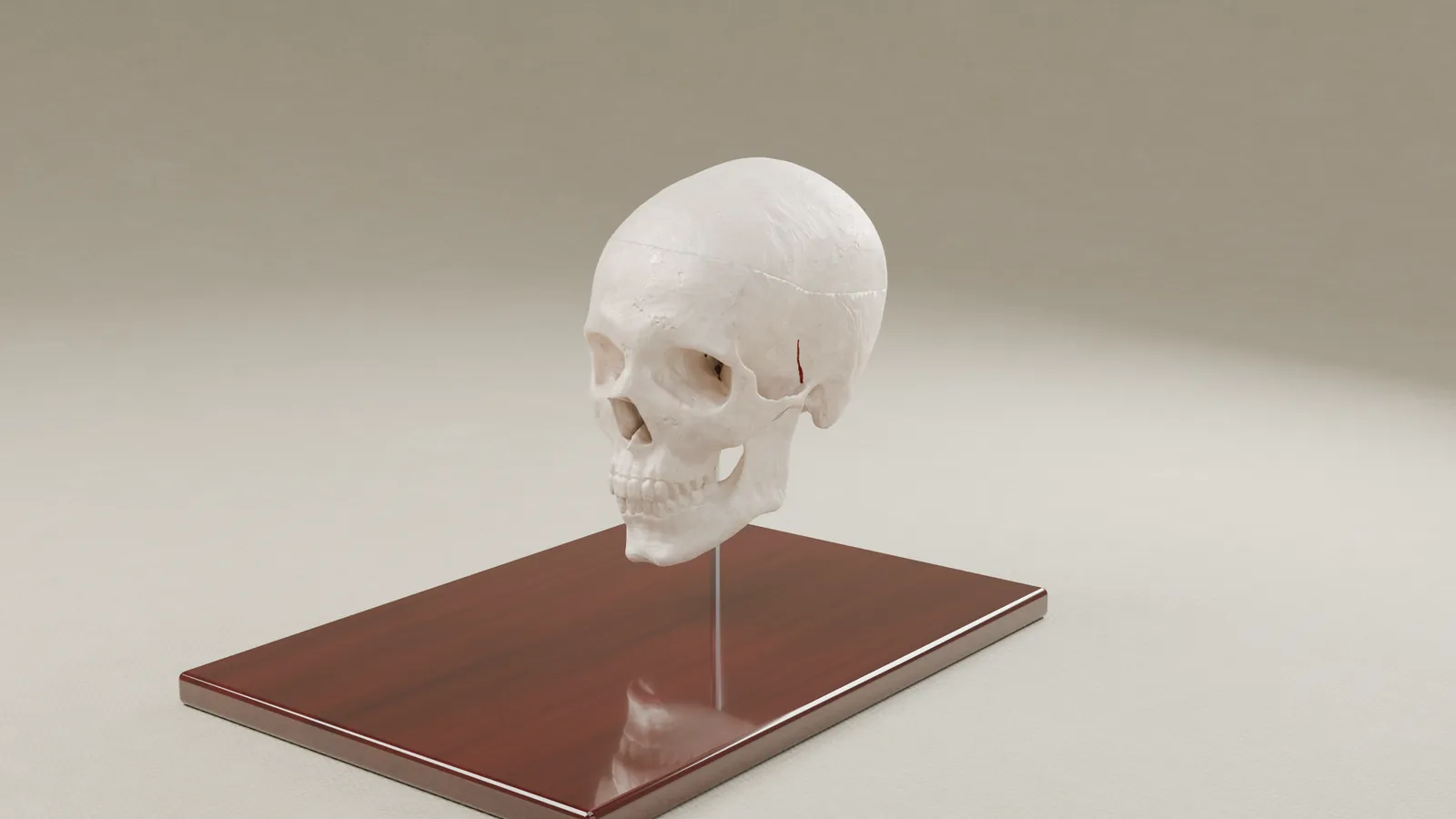
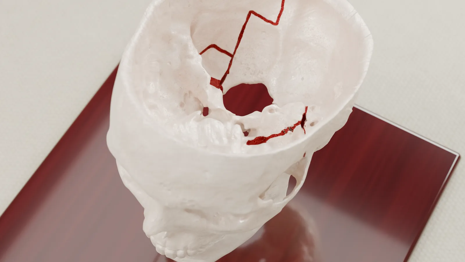
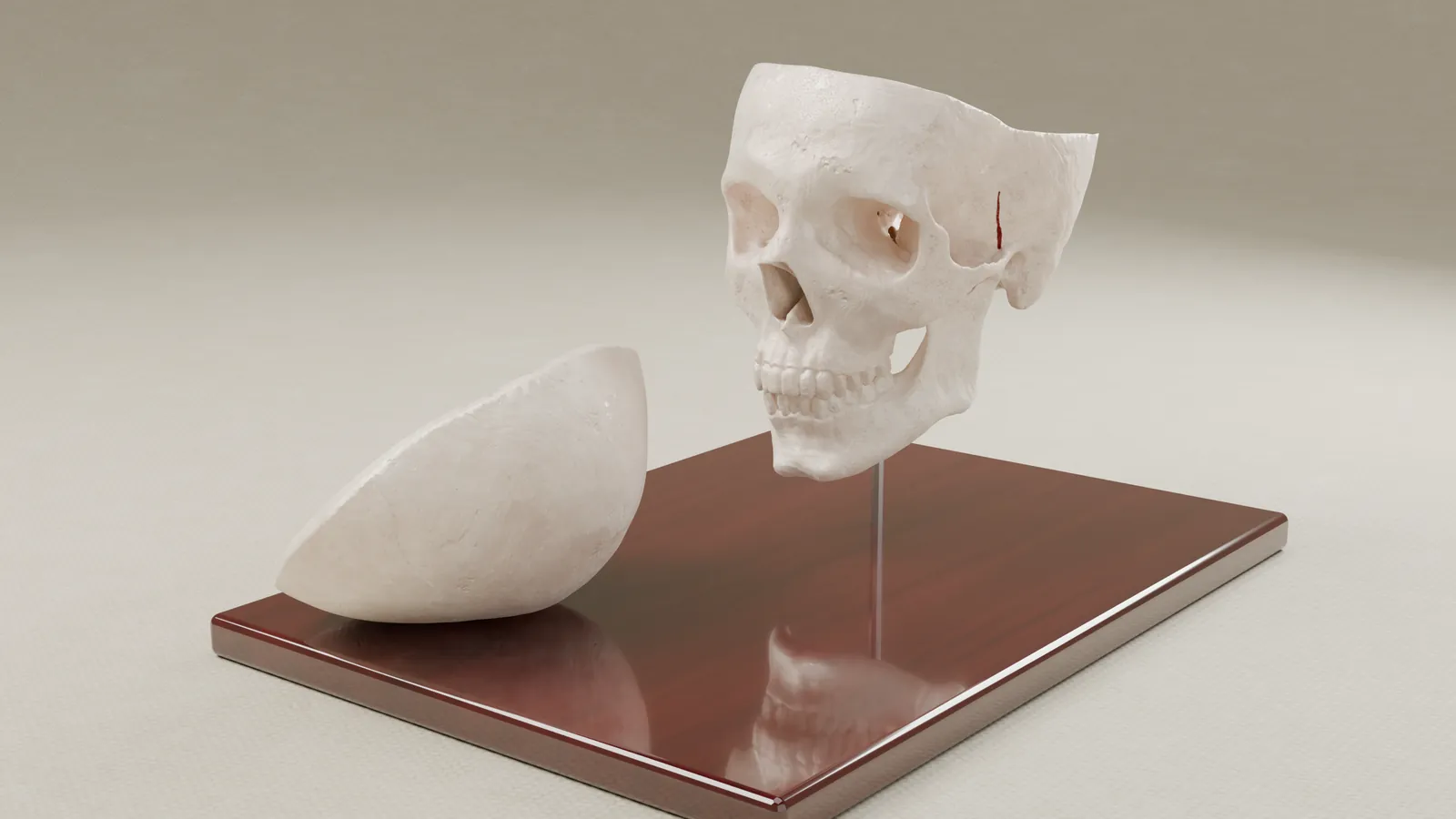
Skull-base fracture in a wrongful death case
For a wrongful death case, we built a split skull from the hospital's own CT scan, showing where the bone was broken without graphic images.
Prior results do not guarantee a similar outcome. Case details are published with client permission; some details are anonymized to protect confidentiality.
Tell us about your case.
Send us the imaging and the question in dispute, and we will tell you what we would build.