Evidence you can see and feel.
From a biomedical engineer, exclusively for plaintiff trials.
For cases that turn on what happened inside the body, we build physical, digital and interactive exhibits from your client's own imaging. You work directly with the biomedical engineer who designs and builds them.
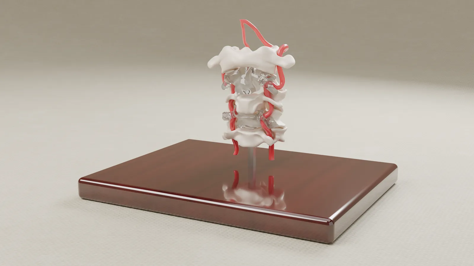
Exhibits built from the case imaging.
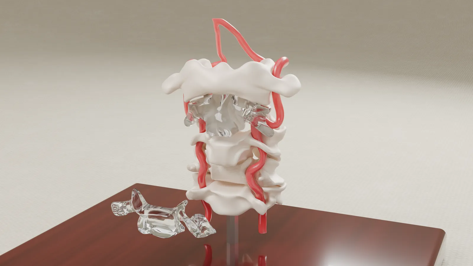
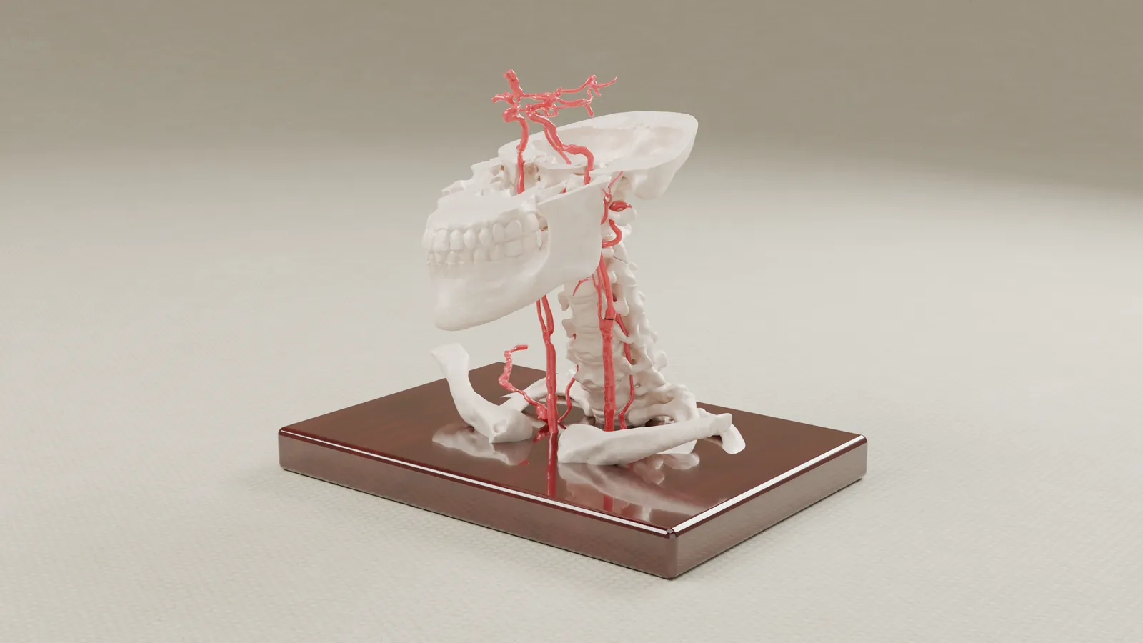
Exhibits for mediation and trial.
Each exhibit is scoped to the facts in dispute and built from the case imaging. Most matters combine more than one of these.
Physical models
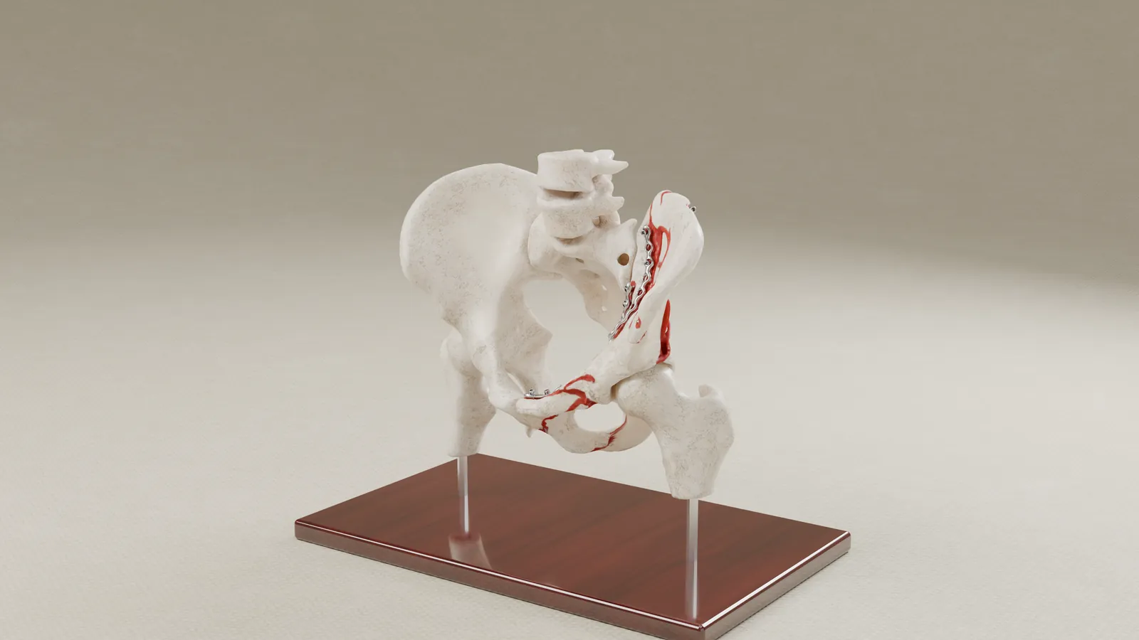
Models of bone and soft tissue built to scale from CT and MRI, including injuries, surgical hardware and scaled replicas of implants. Parts can be transparent or removable, so a jury can see inside the anatomy.
Interactive exhibits
On-screen 3D anatomy that a presenter can rotate, zoom and take apart, with the client's imaging slices shown beside or over the model.
Renderings and animations
Still renderings and short animations made from the same models, for mediation statements, slides and video.
Exhibit planning
We work with counsel and the retained experts to decide what an exhibit has to show, then design it around the facts in dispute.
A specialist partner for medical evidence.
Built from your client's own imaging
Stock models show average anatomy. We build from the case CT and MRI. The exhibit shows this patient, and it can be checked against the source scans.
A biomedical engineer you work with directly
Jim West holds a master's degree in biomedical engineering. You talk to the person who reads the imaging and designs the exhibit, from the first call through delivery.
Plaintiff trials, exclusively
We have worked only for plaintiff attorneys since 2018, so there is no conflict with the firms you face. We work nationwide from the Midwest.
From imaging to delivered exhibit.
Send the imaging and your deadline
Tell us about the matter and what the exhibit needs to show. We review the imaging and the questions in dispute.
We scope the exhibit
We propose what to build and quote it before any work starts.
Counsel approves a digital proof
You review the model on screen and request changes before the final exhibit is built.
We build and deliver
The physical model, interactive exhibit or renderings are built from the approved proof and delivered to counsel.
Before and after, in the client's own anatomy.
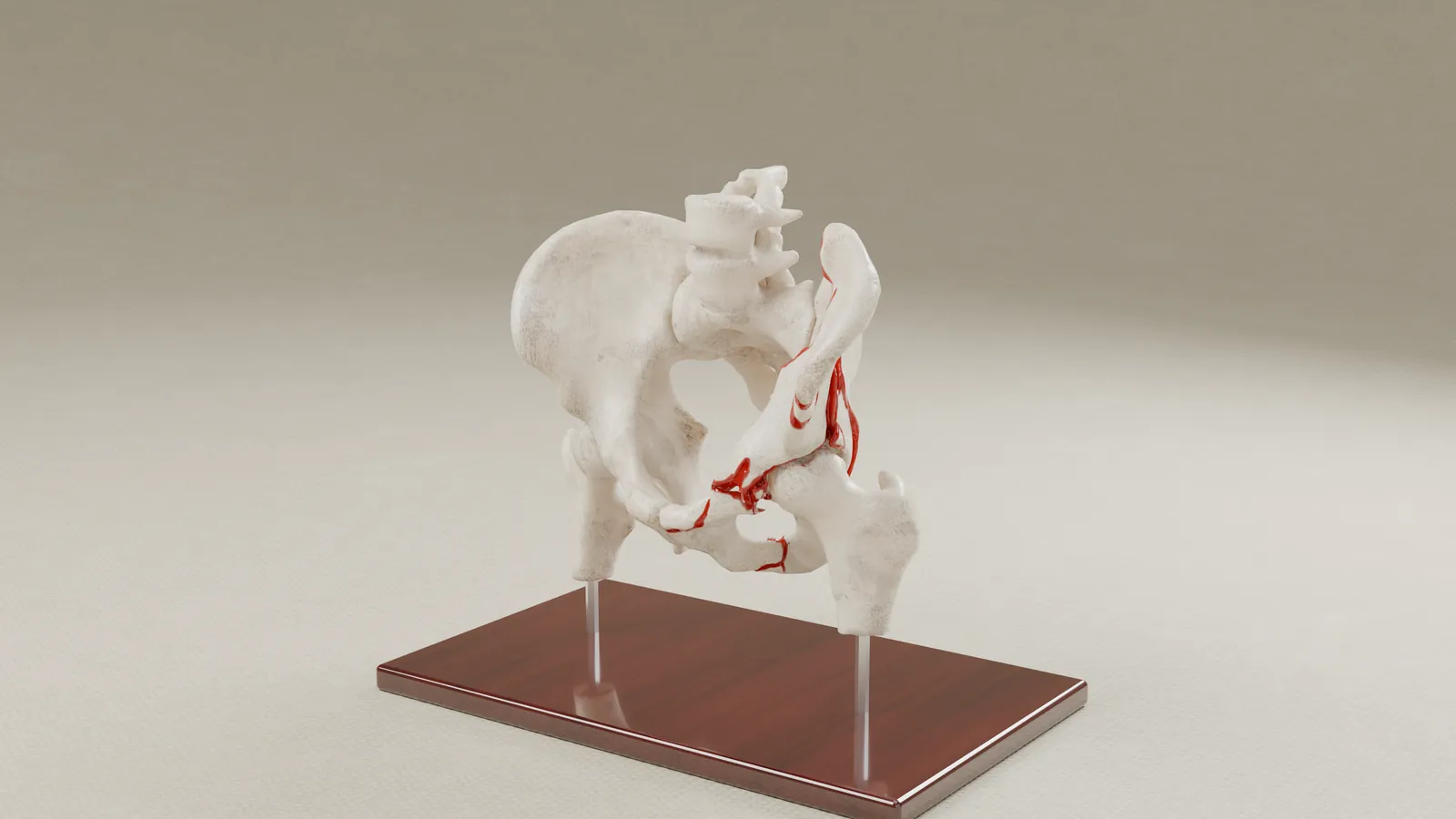
In a pelvic fracture case, we rebuilt the client's pelvis from CT scans taken before and after surgery. The interactive exhibit marks each fracture, moves the plates and screws into their final positions and shows the CT slices over the 3D bones.
"Precision that people can understand."
The principle behind every exhibit we build.
Tell us about your case.
If your case depends on anatomy that is hard to read on a scan, send us a note or book a brief introductory call.