Pelvic fracture and surgical reconstruction: an interactive before-and-after exhibit
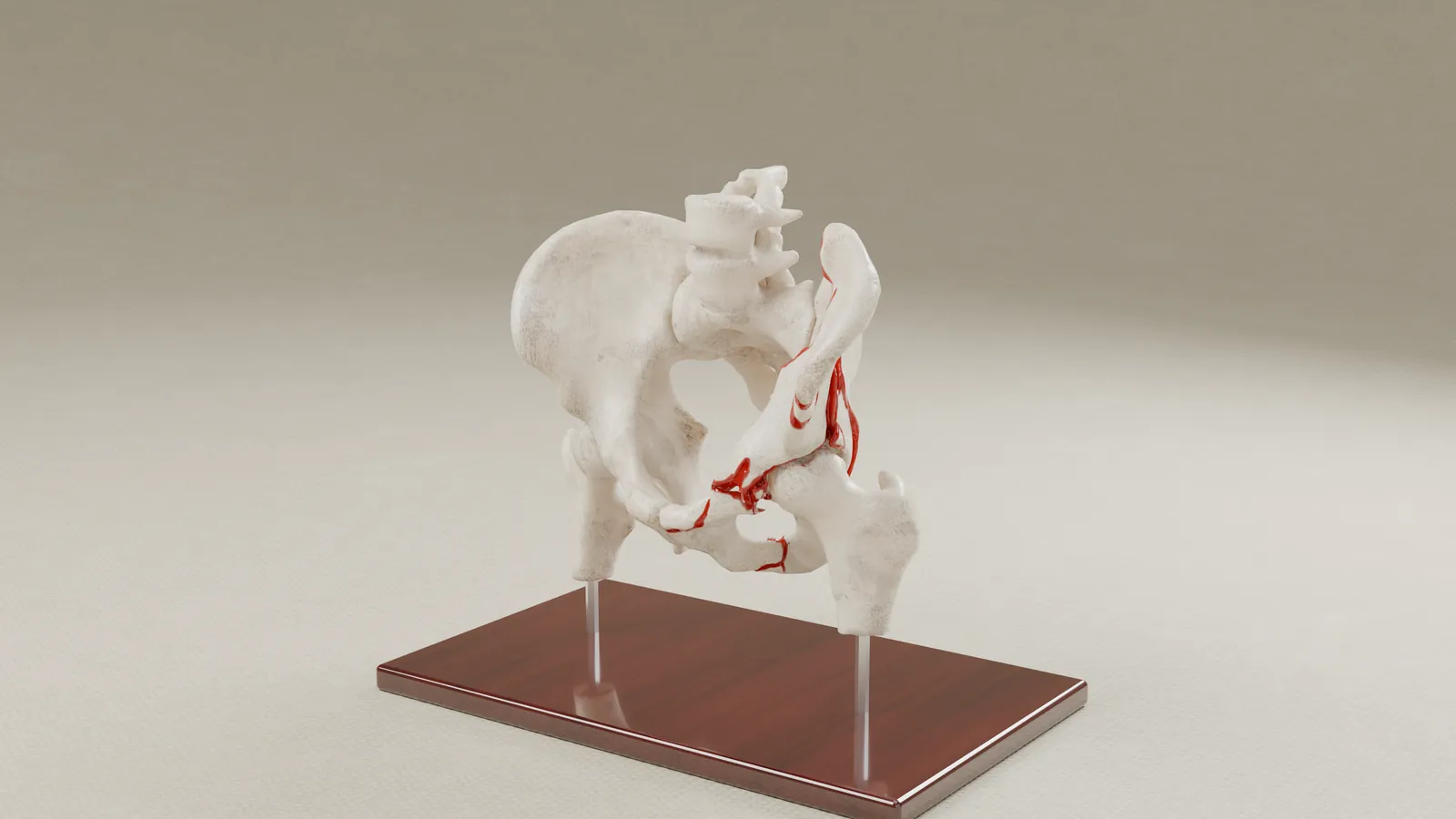
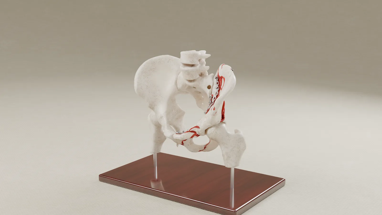
A fall from height fractured a worker's pelvis in several places, and the reconstruction left a large amount of hardware. The firm wanted a mediator and a jury to see both the break and the repair, so we built an interactive exhibit from the client's CT scans, before and after surgery.
- Practice area
- Personal injury
- Anatomy
- Pelvis and hip, fracture and surgical reconstruction
- Client
- Plaintiff-side personal-injury firm
- Exhibit produced
- Interactive digital exhibit, pre-op and post-op, with animated hardware and CT overlay
The challenge
A radiology report can say that a pelvis was fractured and repaired, and illustrations drawn from the report can sketch it. Neither shows how many places the bone broke or how much hardware went in. The firm wanted both states shown in the client's own anatomy, side by side, in front of a mediator well before trial.
The solution
We segmented the client's CT scans from before and after surgery and rebuilt the bones and each piece of fixation hardware. Every bone is kept in its position from the scans, so the two states can be compared directly.
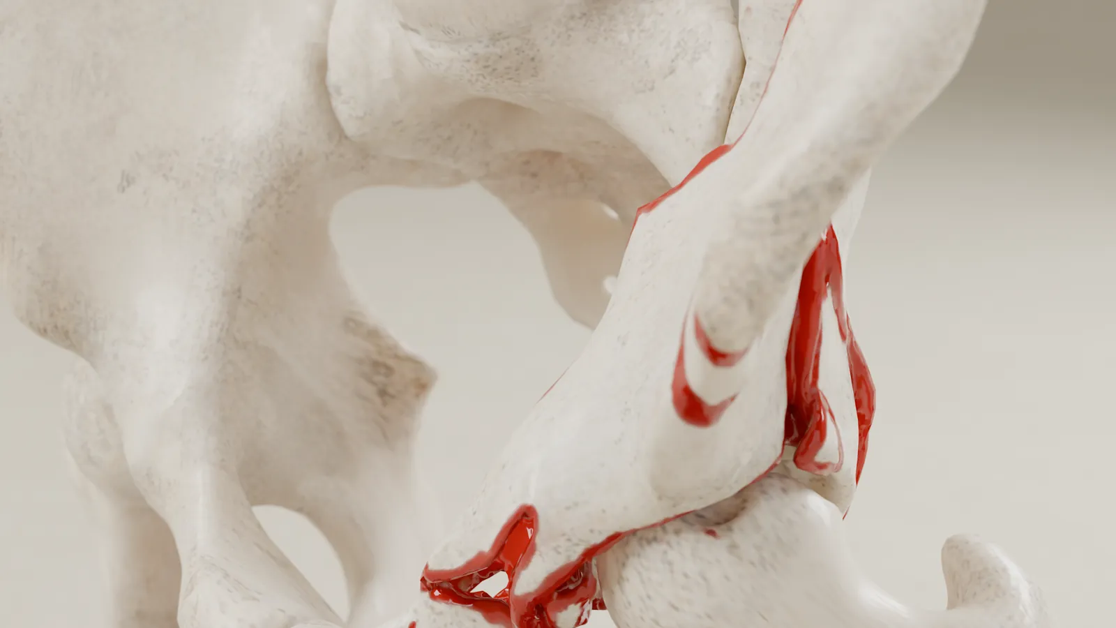
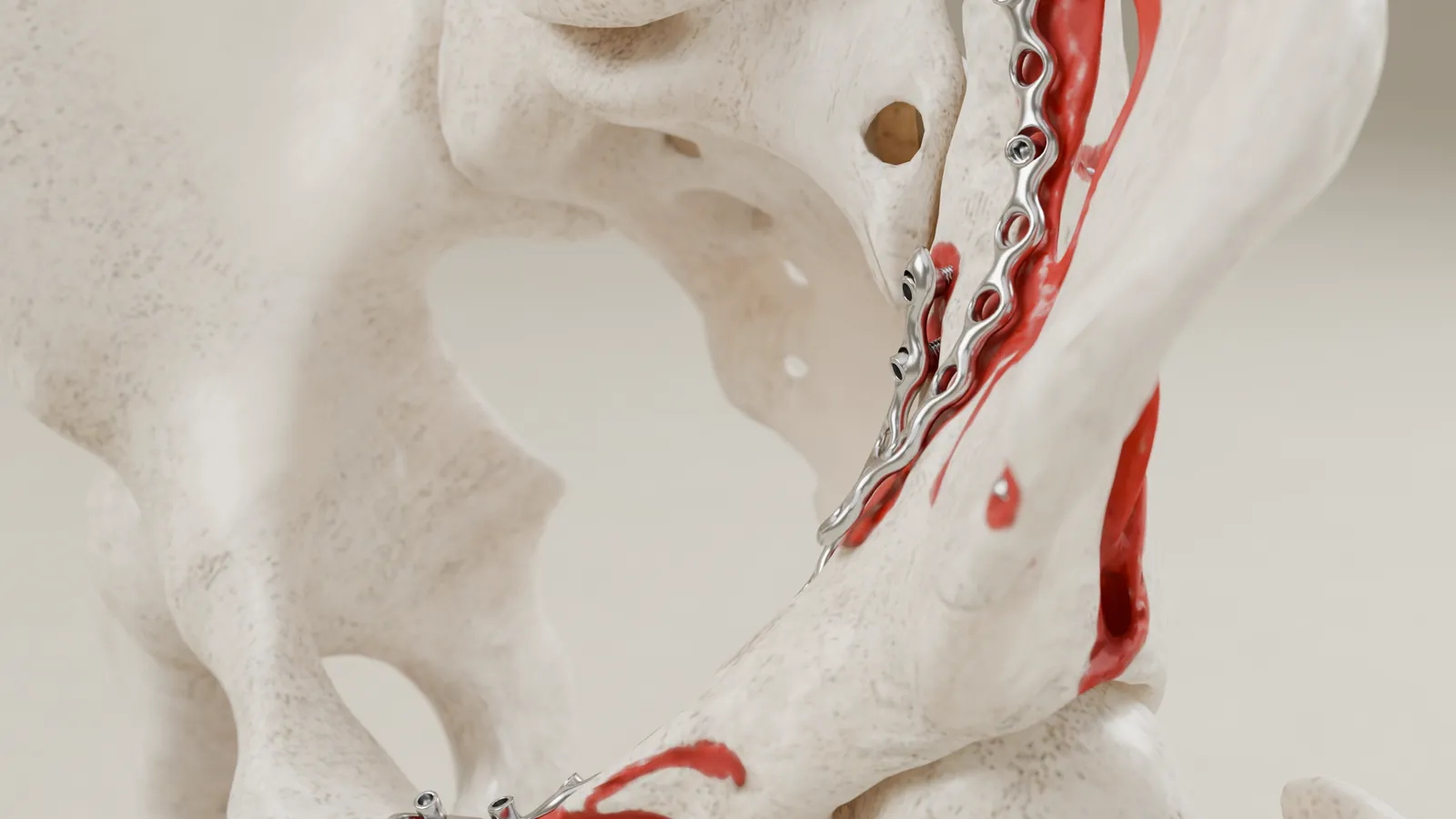
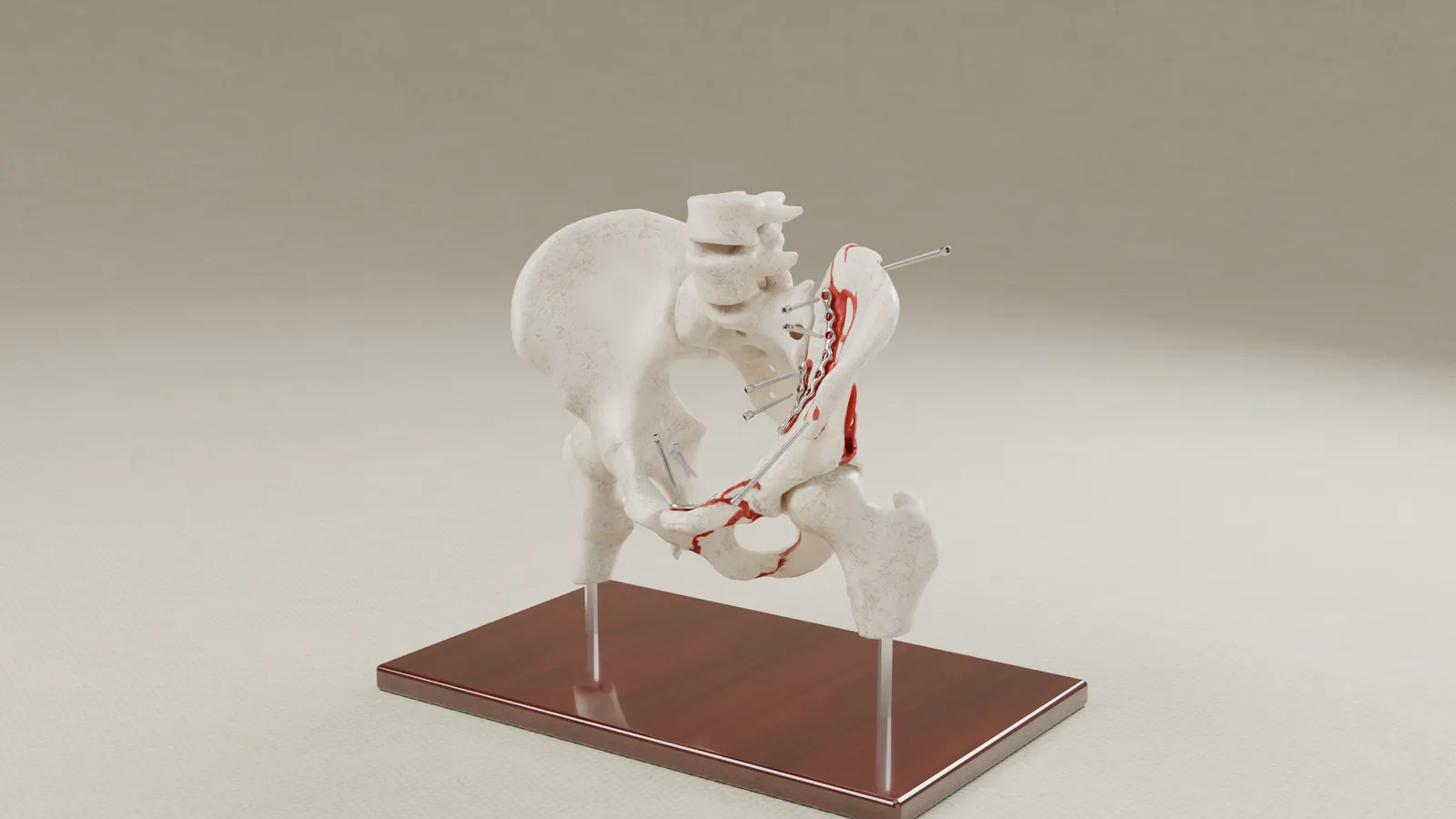
In the interactive exhibit, the fracture lines are marked, the screws and plates move into their final positions on screen, the hardware is labeled, and the actual CT slices can be shown over the 3D bones.
What we built
Interactive exhibit. Two matched models of the client's pelvis, one fractured and one repaired, with marked fracture lines, animated hardware, labels and the CT slices overlaid.
Supporting imagery. Renderings of both models from matching camera positions, plus a view with the screws drawn out of the bone along their own axes.
How it was built
- From the client's own CT scans, before and after surgery.
- Each plate and screw was rebuilt individually from the post-operative scan.
- The exhibit shows the actual CT slices, so the 3D bones can be checked against the scans.
- Built by the biomedical engineer (MS) you work with directly.
If the surgery is part of your damages, send us the scans and we will tell you what we would build.
Prior results do not guarantee a similar outcome. Case details are published with client permission; some details are anonymized to protect confidentiality. Every image on this page is a rendering of the exhibit.