Vertebral artery injury during cervical fusion: a 1:1 model and interactive exhibit
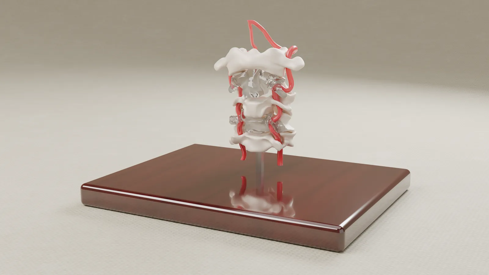
During a planned cervical fusion, a patient's vertebral artery was injured. The plaintiff alleged that the artery's unusual path was visible on the pre-operative MRI. The firm asked us to show that path, so we built a 1:1 physical model and an interactive exhibit from the patient's own imaging.
- Practice area
- Medical malpractice, surgical error
- Anatomy
- Cervical spine, C3-C4, vertebral artery
- Client
- Plaintiff-side medical-malpractice firm
- Exhibits produced
- Physical 1:1 model, interactive digital exhibit with imaging overlay, guided video
The challenge
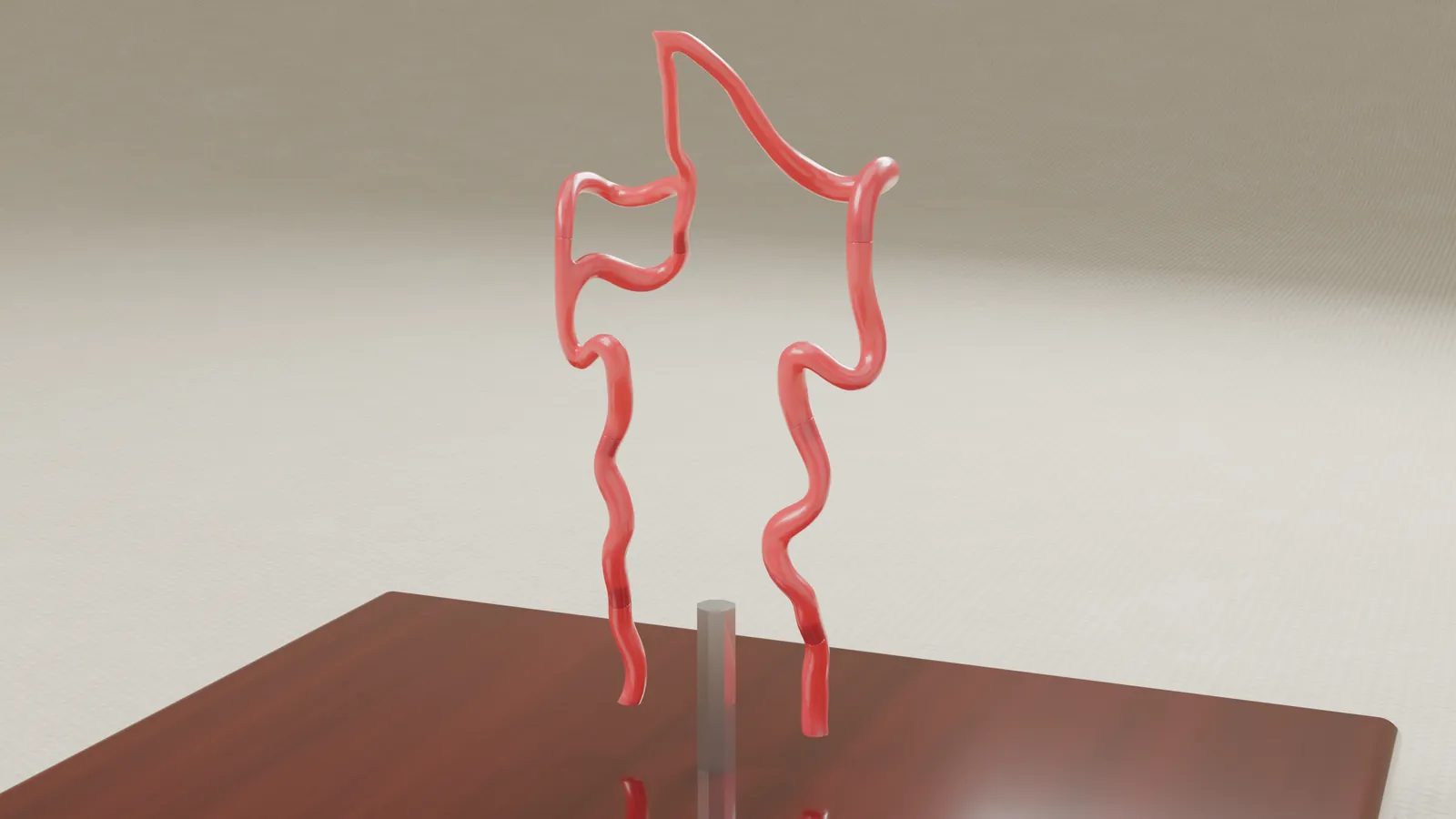
The dispute turned on the path of one vertebral artery as it passes through the small bony channel in each cervical vertebra, and on whether that path could be seen on the imaging taken before surgery. A single MRI slice does not show it. The difference appears only when you scroll through consecutive slices and watch one artery move while the other stays in place. Jurors cannot be expected to rebuild that from flat films, and the firm needed something they could follow without a radiology background.
The solution
We worked from the patient's own CT and MRI data. We segmented the cervical spine and arteries and rebuilt them to scale, showing the anatomy as imaged before the procedure.
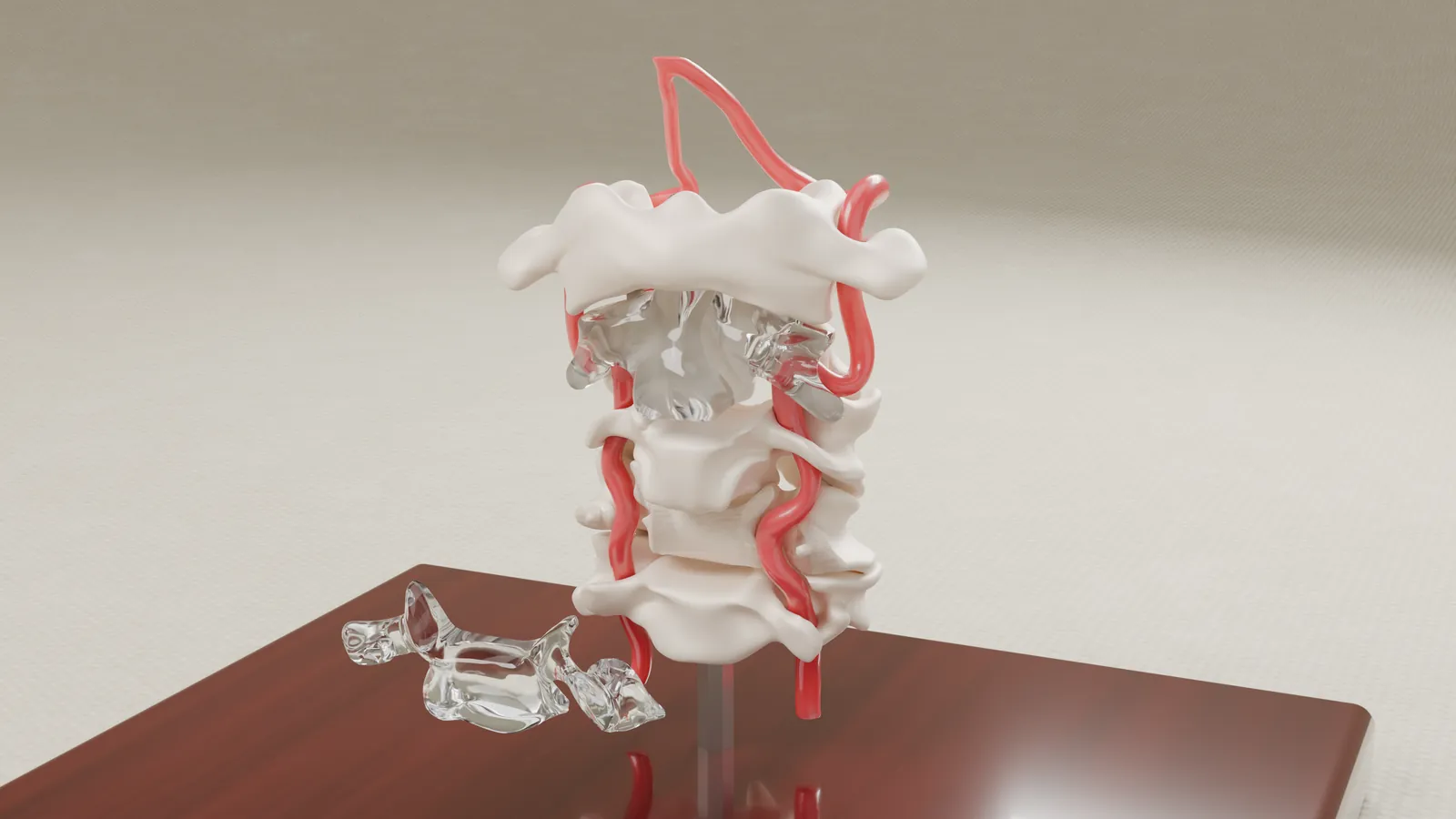
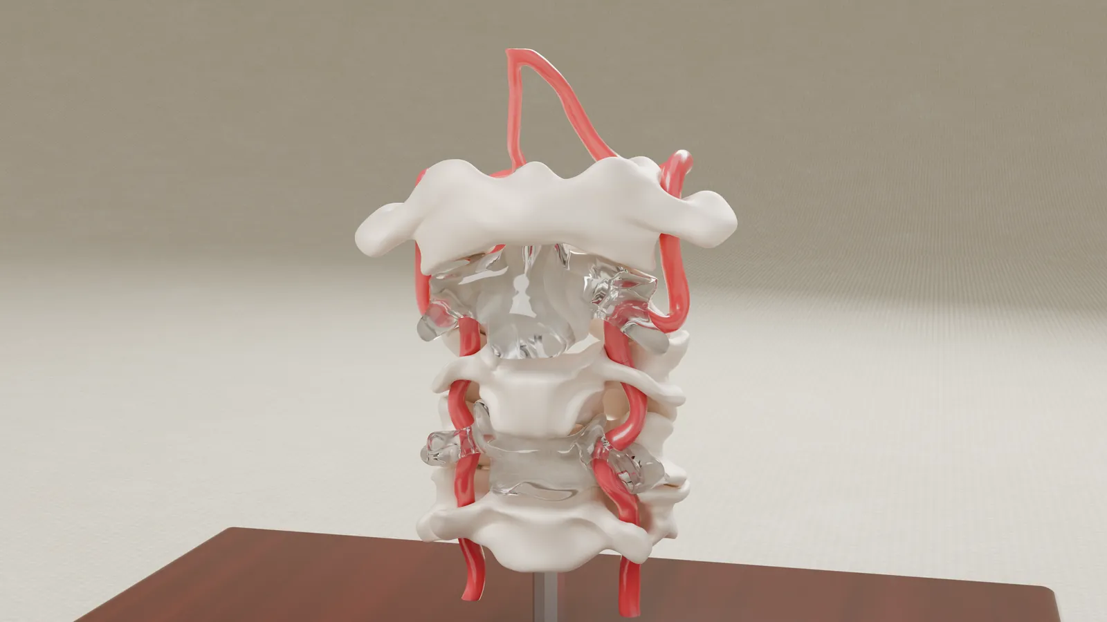
The physical model is 1:1 and holds C1 through C5 above a display base. Some vertebral sections are transparent, so the artery can be seen inside the bone, and one vertebral front lifts off by hand.
The interactive exhibit puts the imaging slices next to the 3D anatomy. A presenter can scroll through the slices, switch imaging layers on and off, change their opacity, move the vertebral fronts out of the way, and label the anatomy.
What we built
Physical model. A 1:1 model of the cervical spine, C1 through C5, with transparent vertebral sections and a front that lifts off to show the artery inside the bone.
Interactive exhibit. The patient's imaging slices beside the 3D model, with imaging layers, adjustable opacity, and animated removal of the vertebral fronts.
Guided video. A walkthrough of the interactive exhibit, made for mediation, settlement and trial preparation.
How it was built
- From the patient's own CT and MRI data, showing the anatomy as imaged before the procedure.
- Scoped with input from the plaintiff's physician expert.
- The model and the exhibit come from the same imaging, so either one can be checked against the source scans.
- Built by the biomedical engineer (MS) you work with directly.
If your case depends on anatomy that is hard to read on a scan, send us the imaging and we will tell you what we would build.
Prior results do not guarantee a similar outcome. Case details are published with client permission; some details are anonymized to protect confidentiality. Every image on this page is a rendering of the exhibit.